Severe impingement (FAI)
The femoral head and socket rim collide during movement, especially when the change extends far towards the back or all around.
Joint-preserving hip surgery
During surgical hip dislocation, the femoral head is lifted out of the socket in a controlled manner during the operation. Its blood vessels are protected throughout. This allows us to inspect the entire joint all around: the femoral head, femoral neck, socket rim, labrum and cartilage. This direct view allows deformities to be corrected completely.
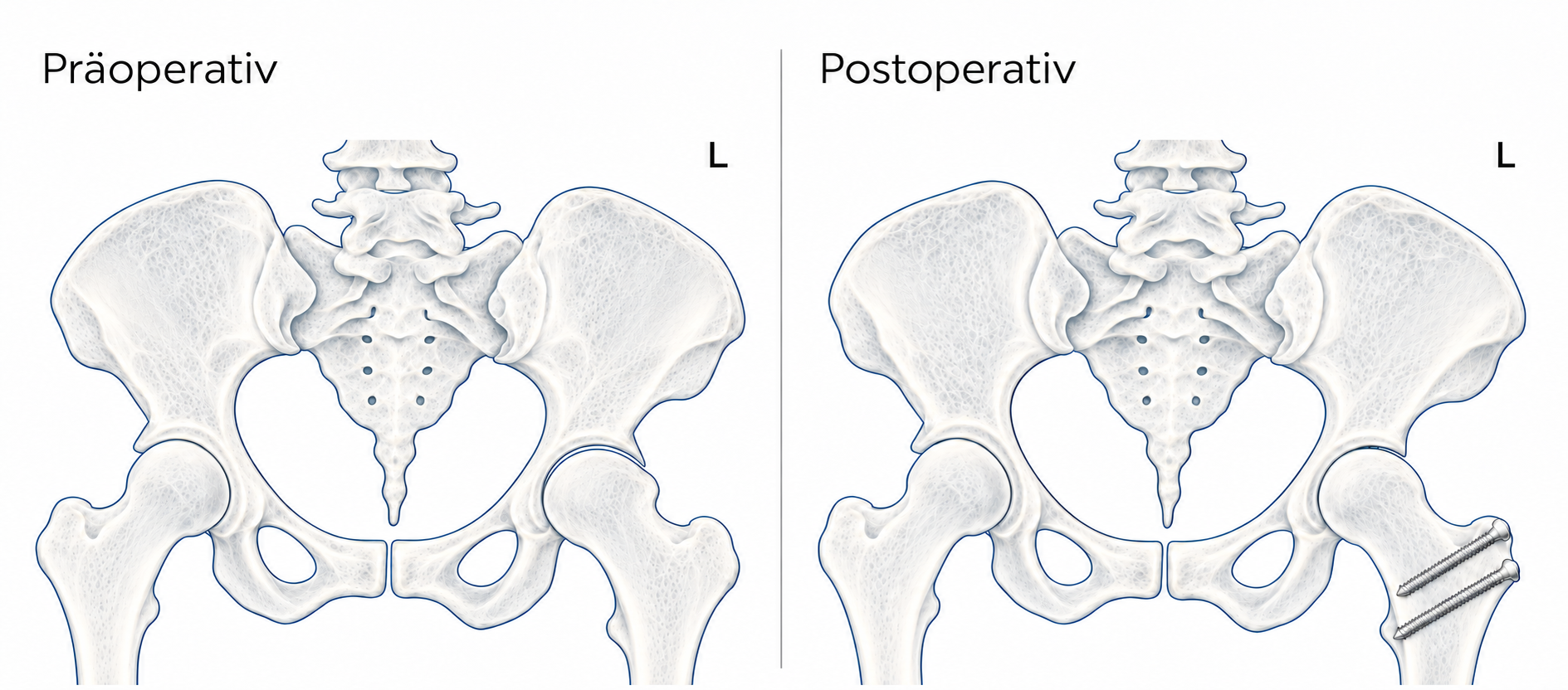
The procedure was developed in Switzerland and has proven itself over many years. It is considered a safe way to treat even complex hip deformities while preserving the joint, meaning without an artificial joint.
We now treat many hip deformities arthroscopically, using keyhole surgery. Surgical hip dislocation may be considered when the change is too extensive or is located in areas that cannot be reached safely with an arthroscope. Typical situations include:
The femoral head and socket rim collide during movement, especially when the change extends far towards the back or all around.
An enlarged or flattened femoral head, a short femoral neck and a high greater trochanter. These lead to pain, limping and restricted movement.
The femoral head slipped during adolescence. This results in a deformity that leads to impingement and early cartilage damage.
The socket rim has become bony or encloses the head too tightly.
For example, benign growths of the joint lining that must be removed completely.
The aim is to remove the cause of wear while the cartilage is still preserved. The earlier the correction is carried out, the better the prospects of preserving your own joint in the long term.
The procedure is performed under general anaesthetic, with a nerve block for pain relief also available if you wish. Depending on its extent, the operation takes approximately two to three hours.
The skin incision is on the outside of the hip. We detach a small piece of bone at the greater trochanter together with the muscles attached to it and temporarily fold it to the side. The muscles themselves are not cut. The joint capsule is then opened in a way that protects the blood vessels to the femoral head. Only then is the head carefully lifted out of the socket.
Depending on your findings, we carry out one or more of these steps:
Before closing, we check the range of movement under direct vision and make sure that nothing is still catching. Finally, the piece of bone at the greater trochanter is securely fixed back in place with two or three screws.
If you also have a hip socket that is too shallow (dysplasia) or a rotational deformity of the thigh bone, surgical hip dislocation can be combined with a pelvic osteotomy (PAO) or a correction of the thigh bone. We discuss this with you individually beforehand.
You will usually stay with us for approximately five to seven days. The day after the operation, you will already get up with our physiotherapists and learn to walk safely with forearm crutches, including on stairs. You will receive a blood-thinning medication to prevent thrombosis. We often also prescribe a medication for a few days to prevent excessive new bone formation in the soft tissues.
The most important thing is that the piece of bone moved at the greater trochanter heals securely. The entire aftercare plan is based on this.
An X-ray check shows whether the bone has healed. If it has, you gradually increase the load up to full weight-bearing and stop using the crutches. Targeted muscle building now begins, especially of the muscles that move the leg out to the side. Outpatient rehabilitation or intensive physiotherapy is useful during this phase.
Your walking pattern returns to normal. Joint-friendly sports such as cycling, swimming or using a cross-trainer are possible again.
Following consultation, more demanding sports involving running, jumping or changes of direction are possible again.
Usually after approximately 4 to 6 weeks
Generally after approximately 3 months
When you can walk safely without crutches; after an operation on the right hip, usually after approximately 6 to 8 weeks
The screws at the greater trochanter can remain in the body. If they cause problems, for example when lying on your side, they can be removed in a small procedure no earlier than approximately one year afterwards.
Surgical hip dislocation is a safe procedure in experienced hands. Nevertheless, as with any operation, there are risks. These include delayed healing of the piece of bone at the greater trochanter, calcification in the soft tissues, thrombosis or wound-healing problems. Thanks to the technique that protects the blood vessels, impaired blood supply to the femoral head is very rare. We discuss all risks with you in detail before the procedure.
Most patients have significantly less pain and better movement after the operation. Many return to sport and work. How good and how lasting the result is depends above all on the condition of the cartilage at the time of the operation. This is why it is worth having symptoms assessed at an early stage.
Appointments & contact
Online, in-person or private consultation: choose the appropriate contact for your appointment. In an emergency, contact the emergency department or call 112. Emails are not monitored around the clock.